Understanding Skin Cancer: Prevention, Detection, and Treatment
Skin cancer is one of the most common forms of cancer worldwide, with rates continuing to rise despite increased awareness. From the frequently occurring basal and squamous cell carcinomas to the more dangerous melanoma, understanding the causes, warning signs, and prevention strategies is essential for everyone. Early detection significantly improves treatment outcomes, making knowledge about skin cancer a potentially life-saving asset.

Skin cancer represents the abnormal growth of skin cells, typically developing on skin exposed to sunlight, though it can occur anywhere on the body. It affects people of all skin tones, though those with lighter skin have a higher risk. With over five million cases diagnosed annually in the United States alone, skin cancer has become a significant public health concern that requires awareness, prevention strategies, and regular screening.
What is skin cancer and how does it develop?
Skin cancer begins when DNA damage in skin cells triggers mutations, causing cells to multiply rapidly and form malignant tumors. This damage most commonly results from ultraviolet (UV) radiation exposure from the sun or tanning beds, though genetic factors and certain environmental exposures also play roles.
The three main types of skin cancer include basal cell carcinoma, squamous cell carcinoma, and melanoma. Basal cell carcinoma affects the basal cells in the lower epidermis and rarely spreads but can be disfiguring if left untreated. Squamous cell carcinoma develops in the middle and outer layers of the skin and has a higher risk of spreading. Melanoma, the most dangerous form, develops from melanocytes (pigment-producing cells) and can spread rapidly to other parts of the body if not detected early.
Other less common types include Merkel cell carcinoma, dermatofibrosarcoma protuberans, and various skin lymphomas. Each type has distinct characteristics, growth patterns, and treatment approaches, highlighting the importance of professional diagnosis.
How to examine moles and recognize changes
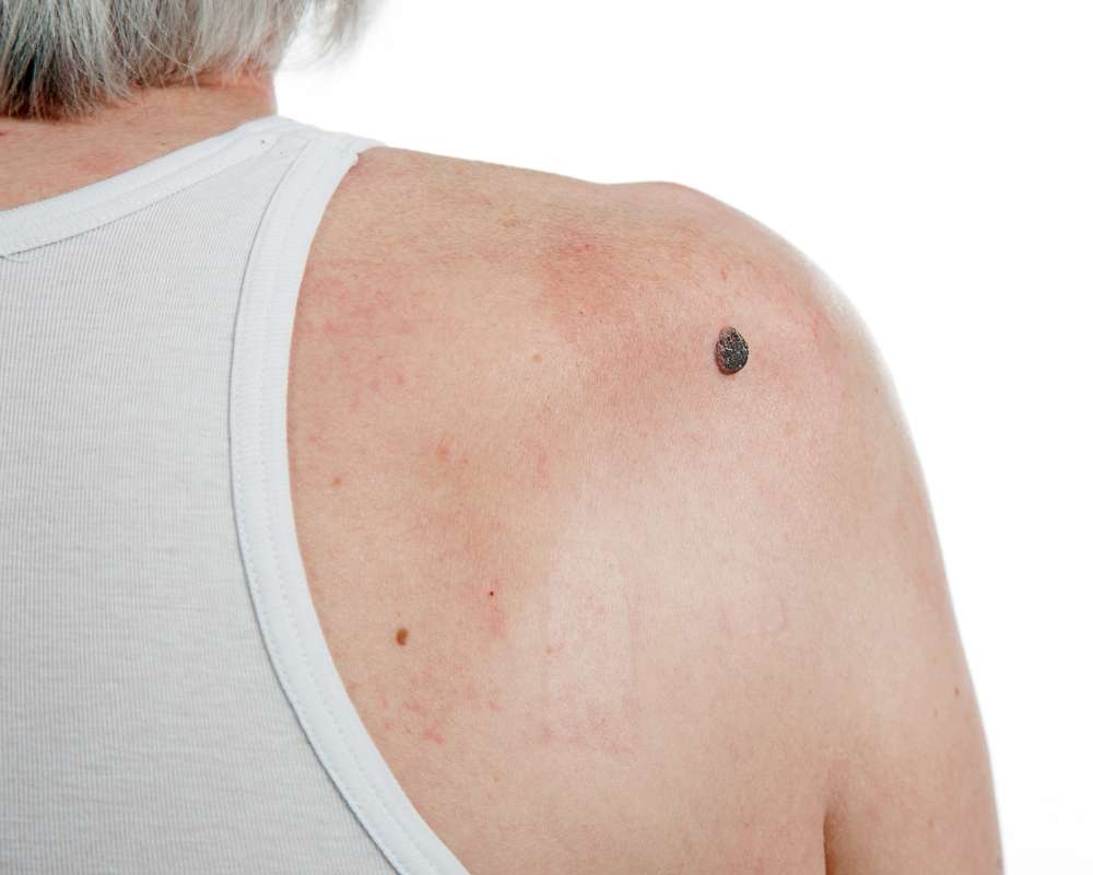
Regular self-examination is crucial for early skin cancer detection. The ABCDE rule provides a helpful framework for evaluating moles:
- Asymmetry: One half of the mole doesn’t match the other half
- Border: Irregular, ragged, notched, or blurred edges
- Color: Variations in color throughout the mole
- Diameter: Larger than 6mm (about the size of a pencil eraser)
- Evolving: Changes in size, shape, color, or elevation
When performing a self-examination, check your entire body, including areas not regularly exposed to the sun. Use mirrors for hard-to-see areas like your back, scalp, and behind your ears. Document suspicious moles with photographs to track changes over time.
Pay special attention to new growths, spots that differ from others, or sores that don’t heal within three weeks. The “ugly duckling” sign—a mole that looks significantly different from others—can also indicate potential problems.
How sunburn relates to skin cancer risk
Sunburn represents acute damage to skin cells from excessive UV radiation exposure. Even a single severe sunburn can significantly increase skin cancer risk, particularly during childhood and adolescence. Research shows that experiencing five or more blistering sunburns between ages 15-20 increases melanoma risk by 80% and non-melanoma skin cancer risk by 68%.
UV radiation damages cellular DNA, creating specific mutations in genes that control skin cell growth. Over time, these mutations accumulate, potentially leading to cancerous growth. Both UVA and UVB rays contribute to this damage, with UVB primarily causing sunburn and direct DNA damage, while UVA penetrates deeper into the skin, generating free radicals that indirectly damage DNA.
The risk is cumulative—each sunburn adds to lifetime damage. People with fair skin, multiple moles, or family history of skin cancer face higher risks, though individuals with darker skin tones can also develop skin cancer, often diagnosed at later stages.
When to consult dermatology for skin concerns
Seeking professional dermatological evaluation is essential in several scenarios. First, schedule an appointment if you notice any of the ABCDE warning signs in moles or skin growths. Additionally, any new growth that bleeds, itches, or doesn’t heal within three weeks warrants examination.
Individuals with risk factors should establish regular screening schedules with a dermatologist. These factors include: - Personal or family history of skin cancer - History of significant sun exposure or multiple sunburns - Fair skin, light eyes, or blond/red hair - More than 50 moles or unusual moles - Weakened immune system - History of radiation therapy
During a skin cancer screening, the dermatologist will conduct a thorough examination of your entire skin surface, possibly using a dermatoscope for closer inspection of suspicious areas. If concerning lesions are found, the doctor may perform a biopsy—removing a small tissue sample for laboratory analysis to determine whether cancer cells are present.
Don’t delay seeking medical attention if you notice concerning changes. Early detection significantly improves treatment outcomes and survival rates, particularly for aggressive forms like melanoma.
Understanding melanoma: signs, diagnosis and prognosis
Melanoma, though less common than other skin cancers, causes the majority of skin cancer deaths due to its aggressive nature and ability to spread rapidly to other organs. It develops when melanocytes—cells that produce the pigment melanin—grow uncontrollably.
Early signs of melanoma often follow the ABCDE rule mentioned earlier. Additionally, melanomas may appear as new, unusual growths or develop within existing moles. They can occur anywhere on the body, including areas not regularly exposed to sunlight, such as between toes, under nails, or on the scalp.
Diagnosis typically involves a skin examination followed by biopsy. If melanoma is confirmed, further tests determine the stage of cancer, which indicates how deeply it has invaded the skin and whether it has spread. Staging ranges from 0 (confined to the epidermis) to IV (spread to distant organs).
The prognosis for melanoma varies significantly based on stage at diagnosis. The five-year survival rate for localized melanoma (Stage 0 or I) exceeds 98%, highlighting the critical importance of early detection. However, once melanoma spreads to distant sites (Stage IV), five-year survival rates drop to about 30%, though newer treatments are improving these outcomes.
Treatment options include surgical removal, immunotherapy, targeted therapy, radiation, and chemotherapy, often used in combination depending on the stage and specific characteristics of the melanoma.
This article is for informational purposes only and should not be considered medical advice. Please consult a qualified healthcare professional for personalized guidance and treatment.


